Scientific Program

Conference Series Ltd invites all the participants across the globe to attend Dentists and Prosthodontics Annual Meeting New Orleans, USA.

Day 1 :

Keynote Forum

James L. Ratcliff

President and CEO Rowpar Pharmaceuticals, USA

Keynote: Evolving concepts of inflammation and oral disease: Have we met the enemy, and is it us?

Time : 10:00-10:30

Conference Series Prosthodontics-2016 International Conference Keynote Speaker James L. Ratcliff photo
Biography:

James L Ratcliff is the Chairman of the Board and CEO of Rowpar Pharmaceuticals, Inc., Scottsdale, AZ, maker of the Dentists Choice and ClōSYS brands of oral care products. In 2014, Rowpar received the US Small Business Administration’s Exporter of the Year Award. From 1989 to 2000, he served as a Senior Research, Scientist, Professor and Director, Center for the Study of Higher Education at the Pennsylvania State University. From 1979 to 1989, he was professor and program head for higher education at Iowa State University. He is the author of 120 articles, books, book chapters, is co-inventor on over 24 patents. He is an active member of the Arizona Biotechnology Assn., American Academy of Oral and Systemic Health, American Education Research Assn., and the International Association for Dental Research. He holds a PhD degree in Higher Education from Washington State University, a MA degree in History from Washington State University, and a BA degree from Utah State University.

Abstract:

There is a chicken and egg question in oral health: Which is the cause of oral disease, the inflammatory response of the host (us) to the insult (invading pathogens, chemicals and treatments), or the pathogens, chemicals and treatments themselves? We know that different people have different inflammatory responses to the same pathogens and chemicals, and we know that systemic diseases, such as adult diabetes, may heighten the inflammatory response to insult. In this presentation, we consider the insult and inflammatory response of certain disease conditions: osteonecrosis of the jaw (BRONJ), lichen planus, and mucositis related to head and neck cancer radiation treatment. So, is it the host’s inflammatory response that is a trigger to disease? And is it the host’s inability to contain microbial overgrowth in the oral microbiome that is a trigger to disease? In short, have we met the enemy, and is it us? Proper home oral care and regular professional visits can overcome our own inability to fully resist disease. Requirements for effective home oral care are reviewed in light of emerging research on sources on insult and inflammatory response.

Keynote Forum

John D Petkanas

Associates In Oro-Facial Pain, LLC, USA

Keynote: Facial trauma and temporo-mandibular disorders

Time : 10:30-11:00

Conference Series Prosthodontics-2016 International Conference Keynote Speaker John D Petkanas photo
Biography:

John D Petkanas has completed his graduation from NewYork University College of Dentistry in 1992 with full academic honours, and then completed his hospital training with clinical affiliates of the Albert Einstein College of Medicine, as Housestaff Chair. He entered private practice in New York City immediately upon completion, and has been practicing in New Jersey since 1998. He has been in clinical practice for over 20 years, and is a Diplomate to both the American Academy of Pain Management and the American Board of Forensic Dentistry. He is a Fellow of the American Academy of Craniofacial Pain, and the American College of Forensic Examiners. His fellowship examination has also been certified by the International College of Craniomandibular Orthopaedics.

Abstract:

Victims of motor vehicle accidents frequently develop cranio mandibular/cranio-cervical dysfunction secondary to their trauma. This should hardly be surprising, as we have reviewed the relevant anatomy and biomechanics involved. Seldom, however, do clinicians such as ER staffers, EMS and first-responders adequately evaluate the temporo-mandibular apparatus for any signs of dysfunction in the immediate post-trauma setting of CAD, unless dislocation is present or fracture suspected. This is not unexpected, as their rightful primary concern is that the patient, our patient, is not going to expire. They are more emergently concerned with cardiovascular stability, integrity of the central nervous system and airway patency. These functions, after all, are primary, central, and essential for life. Neck pains, jaw clicks and low-back strains are secondary, or even tertiary concerns in a trauma setting. This is particularly unfortunate for us because functionally the temporo-mandibular joint, cervical spine and interdental articulations are intimately related, and frequently, problems with these structures may not manifest until sometime has lapsed, status post-MVA. Any alteration in one component must affect the others. Insufficient or delayed diagnosis can allow a long-standing TMD to develop and fulminate into advanced, degenerative temporo-mandibular arthrosis, with devastating results. In light of the above, effective clinical management of all patients suffering cervical hyperextension/hyperflexion injury should include evaluation and treatment of TMJ and masticatory systems function. Clearly then, prompt, aggressive assessment and management of CAD and related craniofacial and temporo-mandibular injuries is vital in order to minimize the effects of said trauma, and to restore the patient to normalcy. To do otherwise is at best negligent and at worst incompetent, as the ramifications can be severe.

  • Track 1: Periodontology Track 2: Cosmetic Dentistry Track 3: Scaling and Root Planing Track 4: Dental Implants
Speaker

Chair

James L. Ratcliff

President and CEO Rowpar Pharmaceuticals, USA

Speaker

Co-Chair

Klenise Paranhos

New York University School of Dentistry, USA

Session Introduction

Klenise Paranhos

New York University School of Dentistry, USA

Title: The effect of collar designs and developments over the years on soft tissue and bone level
Speaker
Biography:

Klenise Paranhos is currently working as a faculty in New York University College of Dentistry, Comprehensive Care Postgraduate International Program, Cariology and Comprehensive Care, New York City, United States.

Abstract:

The integration between implants hard and soft tissues is extremely important and responsible for the success of dental implants. Soft tissues disrupter with consequentially crestal bone loss along the dental implant surface disturbs the prognosis and the success rate of the implant. It is well known to occur when the implant surfaces were exposed and contaminated by the biofilm, generating the marginal tissue inflammation. Different configurations were proposed with the goal of reducing the amount of early MBL (marginal bone level) around the implants, suggesting more stable bone– implant interface. According to the literature, the polished area sub-crestally facilitates higher rates of early MBL, whereas a rough implant surface placed at the bone level reduces the amount bone loss. However, in long term, this crestal bone can be disturbed when the collar get exposed to oral cavity by any degree of inflammation. All these designs introduced to the implant collar may lead to a decreased amount of initial bone loss at the crestal level to prevent the break of the biological seal and consequentially loss of the implant. The question remains as to whether the surface structure on the collar of the implant exposed can be a genuine reason on the majority of implants failures rate. The objective of this presentation is to review a collar design that is available today that can create better biological seal and consequentially preserve the soft tissue health.

Speaker
Biography:

Cumhur Sipahi is currently working as a Professor in the Department of Prosthodontics, Gulhane Military Medical Academy, Ankara, Turkey. He has published many articlespapers in various journals. He has completed his PhD from Department of Prosthodontics, Gulhane Military Medical Academy, Ankara, Turkey.

Abstract:

Purpose: The aim of the present clinical study was to display the benefits of three innovative direct attachment incorporation methods in order to prevent or minimize metal housing debonding from dentures, attachment malpositioning, and acrylic resin entrapment within attachment undercuts in implant supported mandibular overdentures.
 
Methods: Three innovative methods: Pre-coating of metal housings with autopolymerizing acrylic resin (AAR), fabrication of acrylic resin bars for optimal parallelism, and block-out of attachment undercuts with light-cured composite resin onto circumferential plastic spacers were applied to totally 9 patients in order to minimize the incorrect positioning of the attachment components, to prevent debonding of metal housings from the denture and the avoid entrapment of the AAR around the implant-attachment interfaces.
 
Results: Not any acrylic resin entrapment within implant-attachment interface was detected. The positioning of the attachments was correct. After a follow-up period of 1 year, not any debonding of metal housing was seen and all patients used their dentures without any severe complaint. Clinical and radiographic evaluations did not reveal any pathological sign.
 
Conclusion: It was concluded that the 3 innovative direct attachment incorporation methods used in the present study could be used as appropriate alternatives in the attachment incorporation procedures of implant-supported mandibular over-dentures.

Speaker
Biography:

Cumhur Korkmaz is currently working in Gülhane Military Medical Academy, Turkey. He is an Assistant Professor at Gata Haydarpasa Training Hospital and
Assistant Professor of Prosthodontics at the G.A.T.A - Haydar Pasha Training and Research Hospital. Gülhane Military Medical Academy (GMMA) was founded on December 30, 1898 with the contributions of German physicians, Prof. R. Rieder, MD and Prof. G. Deycke, MD, as a modern hospital of the era under the name of Gülhane Military Practice School and Hospital in Istanbul.

Abstract:

Objective: The purpose of this in-vitro study was to compare the marginal fit of hot isostatic pressed (HIP) zirconia copings fabricated 3 different marginal finish lines.
 
Material & Methods: To simulate prepared teeth, 30 stainless steel core specimens were fabricated via CNC machining. Stainless steel specimens, which are 6 mm crown length, 1,2 mm finish lines width and 60-degree axial wall inclination angle, were prepared for comparison. These specimens were prepared and separated 3 different marginal finish lines as shoulder finish line (SHO), chamfer finish line (CHA), bevel finish line (BEV). 30 HIP (hot isostatic pressed) zirconia copings with 3 different finish lines configuration
were fabricated with DCS Precident CAD-CAM System to fit onto stainless steel core specimens.
 
Results: Marginal gap measurements were performed using a stereomicroscope with x150 magnification. Obtained data were analyzed with the SPSS 15.0 statistical software program. Kruskal-Wallis test was used to comparison of multiple groups. The average marginal gap values were obtained as follows, respectively: Group SHO/HIP (35.8±13.5μm) < Group CHA/HIP (44.3±13.3μm) < Group BEV/HIP (55.7±27.6μm). Statistically significant difference was found between groups with different finish line (p<0.001).
 
Conclusion: The amount of marginal gap at hot isostatic pressed (HIP) zirconium frameworks has been affected by different finish line.

Biography:

Seyed Hadi Sajjadi is currently working in X-Ray Lab, Hormozgan Aluminum Corporation, Iran. He has completed his PhD from Islamic Azad University Branch North Tehran in 2009-2011. He is a fellow in Department of Orthodontics, Dental Branch, Islamic Azad University, Tehran, Iran.

Abstract:

Purpose: The effect of image quality or dental specialties on the subjective judgment of facial beauty has not been evaluated in any study. We evaluated the effect of digital sensors, camera lenses and dental specialties on the perception of smile beauty.
 
Materials & Methods: In the first phase of this double-blind clinical trial, 40 female smile photographs (taken from dental students) were evaluated by a panel of 3 prosthodontists, 6 orthodontists, and 3 specialists in restorative dentistry, to select the most beautiful smiles. In the second phase, the 20 students having the most appealing smiles were again photographed in standard conditions, but this time with three different digital sensors: Full-frame 21.1-megapixel, half-frame 18.0-megapixel and compact 10.4-megapixel. The same was repeated with two different lenses: Regular and then macro lenses. The same panel judged smile beauty on a visual analog scale. The referees were blinded of the type of sensors and the images were all coded. The data were analyzed using two-way ANOVA, Kruskal-Wallis and Mann-Whitney U tests (α=0.05, α=0.0167).
 
Results: The mean scores for full-frame, half-frame, and compact sensors were 6.70±1.30, 4.56±1.29, and 4.40±1.39 [out of 10], respectively (Kruskal-Wallis P=0.000). The differences between the full-frame with the other sensors were statistically significant (Mann-Whitney P values<0.01). However, the difference between the half-frame and compact sensors was not statistically significant (P>0.1). The scores given by groups of specialists were not statistically different (Kruskal-Wallis P=0.7). Sensors (ANOVA P=0.0000)
but not specialties (P=0.687) affected the perception of beauty. The lenses led to similar scores of beauty perception (Mann–Whitney P=0.8). There was no difference between subjective beauty perception of specialties (Kruskal–Wallis P=0.6). Two-way ANOVA indicated no significant role for lenses (P=0.1750), specialties (P=0.7677), or their interaction (P=0.7852).
 
Conclusions: The full-frame sensor produced consistently better results and was recommended over half-frame and compact sensors. The photographs taken by a regular lens and then digitally magnified can be as appealing as close-up photographs taken by a macro lens. Dentists of different specialties might have similar standards of smile beauty, although this needs further assessment.

  • Track 5: Sedation dentistry and General Anesthesia Track 6: Treatment modalities Track 7: Periodontal & Prosthodontal Disease Track 8: Dental Implants
Speaker
Biography:

Rebecca Mayall has completed her DDS in 2011 from the University of Tennessee, Health Science Center College of Dentistry in Memphis, Tennessee. After completing her degree, she divided her time between private practice and working as a clinical instructor at the College of Dentistry. In 2013, she was hired as an Assistant Professor in the Prosthodontics Department where she currently lectures in the preclinical courses and instructs in the clinic.

Abstract:

Long-term use of bisphosphonates (BP) is associated with the risk of BP-related osteonecrosis of the jaw (BRONJ). The number of post-menopausal women receiving BP therapy for osteoporosis continues to increase as the life expectancy increases. Dental implant rehabilitations have progressed over the years. The purpose of this study was to evaluate the peri-implant conditions of patients taking oral or intravenous (IV) BPs. This was a retrospective study using an ORACLE database search by means of a Procedural Language/Structured Query engine. Fifty female patients with dental implant rehabilitations and taking bisphosphonates, and fifty female patients with dental implants but not taking BP were evaluated. Peri-implant health and rehabilitation conditions were collected and statistics analyzed. Data analysis revealed a strong indication (p<0.01; 82.81%) of the existence of association between abscesses and the use of bisphosphonates. Relative risk indicated 25 times more chance of a person that does not use bisphosphonates to develop abscess when compared with people using bisphosphonates. This shows that the use of bisphosphonates may play a protecting role in the patients with dental implants. Alternatively, there was a strong indication (p<0.01) of existence of association between the occurrence of mucositis and the use of bisphosphonates. Most of the cases of mucositis (94.44%) occurred in patients that used bisphosphonates while the majority of the cases where mucositis did not occur (59.76%) referred to the group being treated with bisphosphonates. This study revealed that there were not indications of significant associations between doses of bisphosphonates and the occurrence of peri-implant diseases.

Speaker
Biography:

Alper Akgürbüz has completed his PhD from Department of Prosthodontics, Gulhane Military Academy, Medical Academy Department of Prosthodontics. He has attended several international dental congresses and symposiums and presented oral and poster presentations. He is currently working at Etimesgut Military Hospital in Ankara, Turkey.

Abstract:

Treatment planning for missing maxillary lateral incisors is a common clinical predicament encountered by orthodontists and prosthodontists. Three main treatment options exist including: canine substitution; a tooth supported restoration; or a single tooth dental implant. An interdisciplinary approach is important throughout treatment planning, and subsequent treatment can involve dental team members such as an orthodontist, oral and maxillofacial surgeon and prosthodontist. The advantage of using dental implants to replace maxillary lateral incisors lies in excellent success and survival rates, and the lack of need to involve adjacent teeth in a fixed restoration. A 23 year old male patient was referred to the clinic after appropriate orthodontic treatment had been completed. Computerized tomography and clinical examination revealed that two narrow dental implant could be placed for congenitally missing maxillary lateral incisors. Three months after the placement of implants, a modified flap was applied for increasing the buccal soft tissue. Subsequently soft tissue healing, prosthetic restorations were completed with zircone porcelain restoration. The predictability of implant therapy has revolutionized how clinicians treat a wide array of tooth replacement situations, including the problem of a congenitally missing maxillary lateral incisor. Although a number of therapeutic alternatives should be considered, treatment with implants allows the clinician to avoid the preparation of adjacent teeth and provides a predictable and enduring solution to the clinical problem.

Faruk Emir

Gulhane Military Medical Academy, Turkey

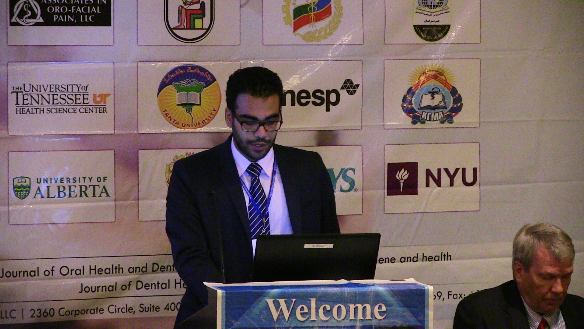
Title: Full-mouth rehabilitation of a patient with severe dental wear
Speaker
Biography:

Faruk Emir has completed his PhD from Department of Prosthodontics, Gulhane Military Medical Academy, Ankara, Turkey. He is working as a Clinical Assistant in Gulhane Military Medical Academy, Department of Prosthodontics. He has published more than 8 papers in various reputed journals.

Abstract:

Severe tooth wear occurs with dental hard tissue destruction due to pathological reasons. Excessive occlusal wear can also cause loss of vertical dimension. In this case after a detailed examination the aim was to recreate the lost occlusal vertical dimension, and to restore the function and esthetics with fixed and removable restorations. Fifty-eight-year-old male patient referred to our clinic with a complaint of impaired esthetics and loss of masticory function. Some of his teeth underwent crown lengthening operation by means of endodontic treatment and prefabricated post along with incrementally built core restorations. Upper and lower classical partial removable prosthesis was fabricated with autopolymerizing acrylic resin material. The occlusion was set according to the canin protected design and followed-up for 2 months with 1-week-interval appointments for the fine re-adjustment of occlusal contacts. After the control of vertical dimension for 2 months final metal-ceramic restorations were fabricated. In the next step maxillary and mandibular removable prosthesis were fabricated and treatment was finished. No complications in our patient and the esthetic, functional and psychological outcomes were all positive 6 months after the end of the treatment. Severe dental wear is compromising for chewing, speaking and esthetic appearance of the dental patients and loss of vertical dimension challenges the treatment. A treatment approach has to be applied with care and patient satisfaction has been achieved in function and aesthetics.

Speaker
Biography:

Klenise Paranhos is currently working as a faculty in New York University College of Dentistry, Comprehensive Care Postgraduate International Program, Cariology and Comprehensive Care, New York City, United States.

Abstract:

The integration between implants hard and soft tissues is extremely important and responsible for the success of dental implants. Soft tissues disrupter with consequentially crestal bone loss along the dental implant surface disturbs the prognosis and the success rate of the implant. It is well known to occur when the implant surfaces were exposed and contaminated by the biofilm, generating the marginal tissue inflammation. Different configurations were proposed with the goal of reducing the amount of early MBL (marginal bone level) around the implants, suggesting more stable bone– implant interface. According to the literature, the polished area sub-crestally facilitates higher rates of early MBL, whereas a rough implant surface placed at the bone level reduces the amount bone loss. However, in long term, this crestal bone can be disturbed when the collar get exposed to oral cavity by any degree of inflammation. All these designs introduced to the implant collar may lead to a decreased amount of initial bone loss at the crestal level to prevent the break of the biological seal and consequentially loss of the implant. The question remains as to whether the surface structure on the collar of the implant exposed can be a genuine reason on the majority of implants failures rate. The objective of this presentation is to review a collar design that is available today that can create better biological seal and consequentially preserve the soft tissue health.

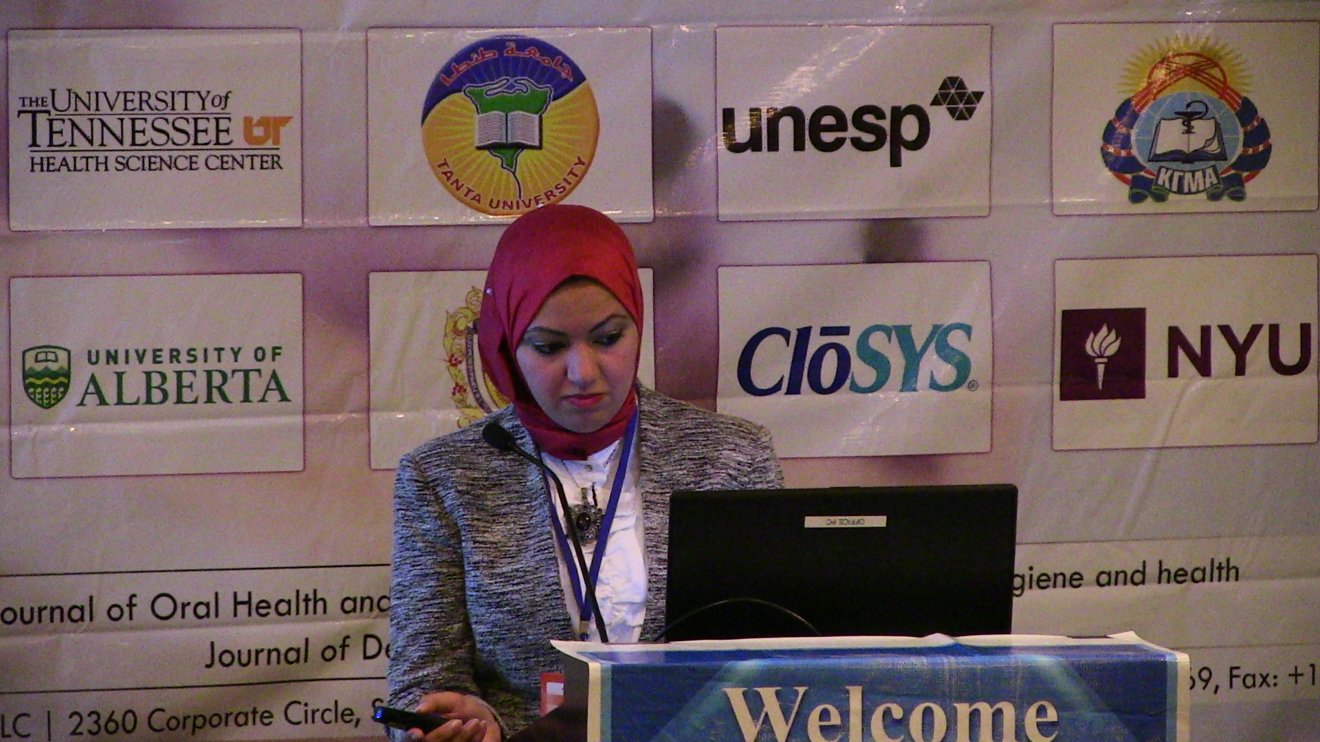
Radwa El-Dessouky

Tanta University Faculty of dentistry, Egypt

Title: To Seek a Longer Conservation Method of Tooth
Speaker
Biography:

Radwa El-Dessouky has completed her MD from Tanta University, Egypt. She works as assistant lecturer at Fixed Prosthodontic Department; Faculty of Dentistry at the same university.

Abstract:

Perfect marginal seal is considered the first defense line of crowned tooth against caries, periodontal disease and hypersensitivity. It was an old concept that metallic margins achieve better adaptability than ceramics and hence metal-ceramic restorations were preferred by both dentists and patients in attempt to combine esthetics, strength and adaptability. All-ceramic crowns are popularly encouraged nowadays thanks to their excellent ability to mimic tooth structure in shade and translucency, biocompatibility and durability. Introduction of newer digital processing generations and ceramic sintering techniques together with the evolution of nano ceramics and resin ceramic hybrid materials are promising issues towards all-ceramic restoration with excellent margins. Additionally, certain details during tooth preparation and laboratorial adjustments were found to have a distinct effect on marginal fit of ceramic restorations. This article provides a brief overview about factors affecting marginal adaptation of ceramic restoration giving more focus on the effect of digital systems, nanoceramics and recent sintering techniques on ceramics marginal fit and longevity in the oral cavity.